|
Since the successful invention of the first Magnetic Resonance Imaging (MRI)
system by Damadian et al. for cancer diagnosis three decades ago, the medical use of
MRI has developed rapidly applicable to a wide range of diseases. Since the first
attempt to image the heart with MRI in the early 1980s, extensive hardware and software
advance had overcome the initial obstacles of gating a rapidly beating heart and
long acquisition times. Over the past years, cardiovascular MRI has become a crucial
tool in many routine clinical cardiac diagnoses, with the advantage of providing not
only structural but a multitude of functional or physiologic data which are adjunctive or
even superior to conventional imaging tools.
Cardiovascular Magnetic Resonance Imaging comprises the efforts of a team of
international authors from different disciplines who contributed a broad range of expertise
to the current field of cardiovascular MRI. Throughout the production of this book, the
emphasis has been on comprehensive, scientifically accurate, and clear explanations of
the many components of this rapidly advancing field. We aim to maintain a balance
between technical basis, cardiac physiology, clinical validation, and available prognostic
implications to enhance the educational value of this reference textbook. Radiologists,
cardiologists, internists, and residents and fellows with interests in cardiovascular MRI
may benefit from the range of materials discussed in this book. The accompanying CD
aims to provide an improved interactive learning experience by including many clinical
case presentations.
While plenty remains to be explored in unleashing the full potential of magnetic
resonance technology in imaging the heart, the field of cardiovascular MRI has experienced
steady growth with balanced emphases in both technical improvement and
clinical application. The first edition of the Cardiovascular Magnetic Resonance
Imaging is intended to serve as a practical introductory resource in helping our readers
overcomes the starting challenges in this field.
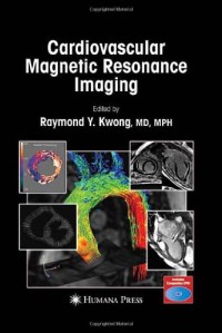
To better illustrate the versatility of cardiac MRI, we designed the front cover of
this book to include multiple cardiac MRI techniques. Going in a counter-clockwise
fashion: cine steady-state free precession imaging in a patient with severe mitral
regurgitation; delayed enhancement imaging of myocardial infarction with the heart
contained in a restraint device in a large animal; quantitative analysis of regional left
ventricular function by displacement encoding stimulating echo technique (DENSE);
and a 3D high-resolution visualization of the ascending aortic blood flow in a normal
subject (courtesy of Drs. Michael Jerosen-Herold, FrederickChen, Anthony Aletras,
and Michael Markl, respectively).
I wish to express my appreciation to the editors at Humana Press for the opportunity to
undertake this project and for their outstanding help and support in bringing
Cardiovascular Magnetic Resonance Imaging to fruition. I am especially grateful to my
mentors and colleagues around the world in the field of magnetic resonance imaging who
have contributed these important chapters. I hope you would enjoy reading this book. |